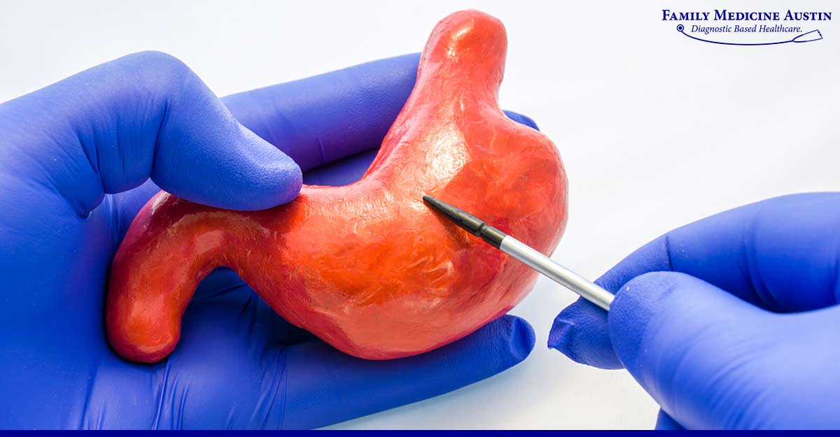
Bleeding in the upper gastrointestinal tract is a common medical issue that physicians encounter frequently. Hematemesis is a frequent sign of this condition, characterized by vomiting blood or a substance like coffee grounds. The condition is also present with melena, i.e., black, tarry stools. In severe cases, the common symptom is hematochezia, i.e., rectal bleeding.
When a patient is suspected of having upper gastrointestinal tract bleeding, the initial examination consists of checking the patient’s blood pressure, searching for relevant risk factors, and determining the care. An endoscopic examination can be performed to determine the cause of the bleeding.
Today’s post presents the pathophysiology of GI bleeding and treatment options to manage the issue. So, continue reading to learn about GI bleeding pathophysiology.
Bleeding in the upper gastrointestinal tract refers to bleeding that originates from the esophagus, stomach, or duodenum (small intestine). It is a common medical emergency with symptoms including anemia, blood or coffee-ground-like material vomiting, black tarry stools, and abdominal pain. Hypovolemic shock may occur in extreme circumstances, resulting in organ failure and death.
Bleeding in the upper gastrointestinal tract is caused by various disorders, including peptic ulcers, gastritis, diverticulitis, and malignancy. The pathophysiology of gastrointestinal (GI) bleeding involves disrupting the blood arteries that supply the GI tract, resulting in bleeding.
Conditions that are associated with the pathophysiology of GI bleeds are discussed below:
The majority of GI bleeds are caused by stomach and duodenal ulcers. Persons with peptic ulcers exhibit bleeding in the upper gastrointestinal tract as their primary symptom. Duodenal ulcers are four times as likely than stomach ulcers to cause bleeding. The proximity of posterior duodenal ulcers to GDA branches makes them more likely to hemorrhage than other duodenal ulcers.
Helicobacter pylori are responsible for the majority of cases of peptic ulcers. H. pylori is frequently associated with persistent and long-term bleeding in the upper gastrointestinal tract. Long-term usage of over-the-counter medications and nonsteroidal anti-inflammatory drugs (NSAIDs) can also induce peptic ulcers. New medications, such as H2 blockers and proton pump inhibitors, which prevent the body from producing acid, have significantly advanced the treatment for peptic ulcers. However, these individuals are more likely to experience rebound or an increase in acid secretion after abruptly discontinuing the medication. Thus, it is essential to inquire about medical history.
Stress ulcers can result in multisystem trauma, hypotension, respiratory failure, sepsis, and jaundice. It may be caused by bile reflux, which damages the stomach’s protective barrier, or by splanchnic vasoconstriction, which restricts blood supply to the liver. Acute gastroduodenal lesions may result from a shock, an infection, surgery, trauma, burns, or a brain condition that leads to GI bleeding.
One-third of all upper GI bleeding is caused by diffuse gastritis. The condition is characterized by several erosions, with the majority occurring in the fundus and body of the stomach. NSAIDs, alcohol, and steroids increase the likelihood of bruising since they are detrimental to the stomach lining. H. pylori is also associated with slow, protracted bleeding.
Varices are enlarged veins in the submucosa caused by increased pressure in the portal vein. Varix ulceration, which can be brought on by reflux esophagitis or increased pressure within varix, is the initial stage in the path to variceal bleeding. Variceal bleeding is responsible for upper gastrointestinal bleeding in cirrhosis and portal hypertension patients. These bleeds pose a threat to the patient’s life. Patients with liver illness produce fewer clotting factors, which increases the likelihood that bleeding will cause complications. Knowing the severity of liver illness is crucial to provide better care.
Dieulafoy’s lesions are large, intertwining blood arterioles in the submucosa of the stomach. Most lesions occur in the fundus and body of the stomach, along the stomach’s slight curve. Since there is a hole in the gastric mucosa, Dieulafoy’s lesions induce bleeding. This hole results from pressure exerted by the bulging and pulsing arteriole.
Both malignant and benign cancers of the upper gastrointestinal tract can produce bleeding. Neoplasms are known to induce light and consistent bleeding, and patients frequently exhibit symptoms of anemia. Endoscopy and biopsies are typically used to determine what is wrong with these tumors.
Aortoenteric fistulas occur when a prosthetic graft in a patient who has had aortic repair degrades into the intestine due to an infection surrounding the graft. An abdominal aortic aneurysm pressing against the colon caused the bleeding. Patients frequently experience a little bleed that resolves on its own, followed by a massive bleed that causes their blood pressure to drop rapidly and need immediate medical attention.

The treatment of GI bleed is based on the bleeding severity and the underlying cause. The initial evaluation consists of a comprehensive patient history, physical examination, and diagnostic tests to identify blood loss and the patient’s overall health status. Diagnostic imaging techniques such as upper gastrointestinal endoscopy, colonoscopy, and radiographic examinations may be performed to determine the source of bleeding.
The following steps may be used to manage GI bleed pathophysiology:
GI bleed management necessitates a multidisciplinary strategy comprising gastroenterologists, surgeons, and critical care specialists to maximize outcomes and reduce complications.
Bleeding in the upper gastrointestinal tract is a medical emergency that, if not treated quickly, might be fatal. The blood arteries supplying the esophagus, stomach, and duodenum are damaged in the pathophysiology of upper GI bleeding, which results in hemorrhage.
A multidisciplinary strategy is used to treat upper GI bleeding, including resuscitation, locating the cause, and administering the proper medications. For identifying and treating upper GI bleeding, endoscopy is frequently the first line of treatment; however, in more serious situations, angiography, embolization, or surgery may be necessary.
Patients with upper GI bleeding receive comprehensive care from GI specialists at Family Medicine Austin. Many occurrences of upper GI bleeding can be successfully treated, and the risk of consequences is reduced with prompt diagnosis and therapy.
Get medical help immediately if you or a loved one exhibits upper GI bleeding symptoms. Contact us for an assessment and treatment. You can regain your health and avoid more issues with the appropriate therapy.